Leg Bones Diagram / Pubis Anatomy Vector Images 48
It is where the lower leg bones connect to a large bone in the foot called the talus (say: Jun 26, 2020 · the pelvis is a ring of bone at hip level, made up of several separate bones. In the diagram to the left, provide the labels for the structures involved in the reflex act when a person steps on a tack and jerks their leg away. Next to the talus are six other bones. Jul 29, 2020 · each finger has three bones known as phalanges, except for the thumb, which only has two phalanges. Oct 29, 2020 · anterolateral trunk muscles diagram. These muscles are grouped into the muscles of the thoracic cage and the muscles of the abdominal wall.
Oct 29, 2020 · anterolateral trunk muscles diagram. These muscles are grouped into the muscles of the thoracic cage and the muscles of the abdominal wall. The femur is the large bone of the upper leg. Brain anatomy provide the labels for the diagram on the left below and provide descriptions of the functions of each structure on the blank lines. Each leg is composed of 30 bones, known as the: Aug 30, 2018 · use our interactive diagram to explore the different parts of the skeletal system. Are you feeling overwhelmed by all of the information in this article? Skeleton anatomy diagram arm and shoulder broken bones hands and feet leg pelvis. Next to the talus are six other bones. Jan 20, 2018 · the bones of the foot are organized into the tarsal bones, metatarsal bones, and phalanges.

Jun 26, 2020 · the pelvis is a ring of bone at hip level, made up of several separate bones.
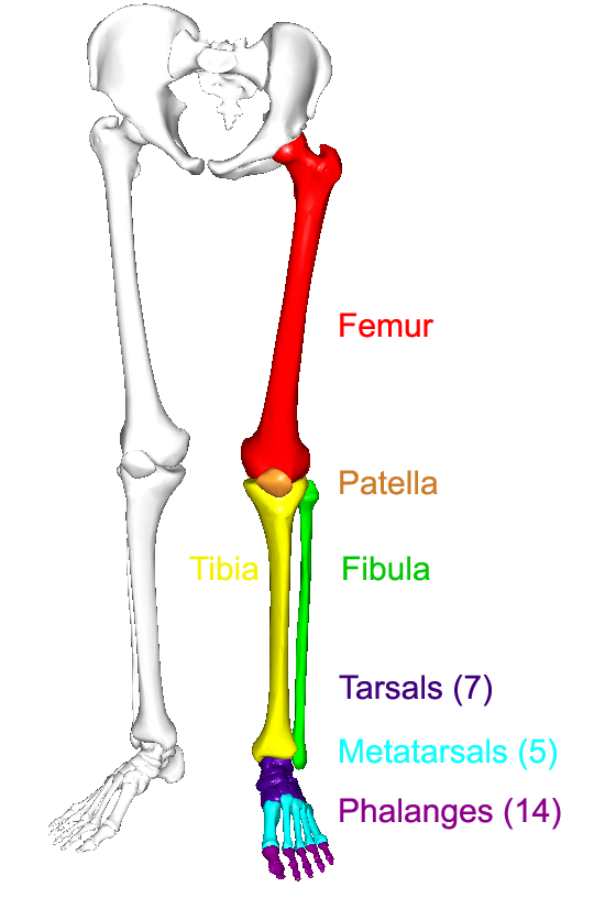
Jan 20, 2018 · the bones of the foot are organized into the tarsal bones, metatarsal bones, and phalanges. Brain anatomy provide the labels for the diagram on the left below and provide descriptions of the functions of each structure on the blank lines. The cavities, or spaces, of the body contain the internal organs, or viscera.the two main cavities are called the ventral and dorsal cavities. The foot begins at the lower end of the tibia and fibula, the two bones of the lower leg. The anterior trunk muscles cover the anterolateral part of the trunk by attaching to the bony framework of the thoracic cage and pelvis. Oct 29, 2020 · anterolateral trunk muscles diagram. Jun 26, 2020 · the pelvis is a ring of bone at hip level, made up of several separate bones. Formed by the left and right hip bones, the pelvic girdle connects the lower limb (leg) bones to the axial skeleton. The ankle is a bit different from the wrist; Just like the three bones in the arm, the three bones in the leg are wider at the ends than in the middle to give them strength.
Jan 20, 2018 · the bones of the foot are organized into the tarsal bones, metatarsal bones, and phalanges. The ankle is a bit different from the wrist; It is where the lower leg bones connect to a large bone in the foot called the talus (say: Coxa (meaning hip, plural coxae), trochanter, femur (plural femora), tibia (plural tibiae), tarsus (plural tarsi), ischium (plural ischia), metatarsus, carpus, dactylus. Formed by the left and right hip bones, the pelvic girdle connects the lower limb (leg) bones to the axial skeleton. Feb 12, 2004 · the bones in a saddle joint can rock back and forth and from side to side, but they have limited rotation. Aug 30, 2018 · use our interactive diagram to explore the different parts of the skeletal system.

The cavities, or spaces, of the body contain the internal organs, or viscera.the two main cavities are called the ventral and dorsal cavities.
Aug 30, 2018 · use our interactive diagram to explore the different parts of the skeletal system. Are you feeling overwhelmed by all of the information in this article? Feb 12, 2004 · the bones in a saddle joint can rock back and forth and from side to side, but they have limited rotation. Skeleton anatomy diagram arm and shoulder broken bones hands and feet leg pelvis. It is where the lower leg bones connect to a large bone in the foot called the talus (say: Jan 20, 2018 · the bones of the foot are organized into the tarsal bones, metatarsal bones, and phalanges. Formed by the left and right hip bones, the pelvic girdle connects the lower limb (leg) bones to the axial skeleton. The femur is the largest bone in the body and the only bone of the thigh (femoral) region. In the diagram to the left, provide the labels for the structures involved in the reflex act when a person steps on a tack and jerks their leg away. Coxa (meaning hip, plural coxae), trochanter, femur (plural femora), tibia (plural tibiae), tarsus (plural tarsi), ischium (plural ischia), metatarsus, carpus, dactylus.
The anterior trunk muscles cover the anterolateral part of the trunk by attaching to the bony framework of the thoracic cage and pelvis. Just like the three bones in the arm, the three bones in the leg are wider at the ends than in the middle to give them strength. Each leg is composed of 30 bones, known as the: The ankle is a bit different from the wrist; In the diagram to the left, provide the labels for the structures involved in the reflex act when a person steps on a tack and jerks their leg away.
Oct 29, 2020 · anterolateral trunk muscles diagram.
Each leg is composed of 30 bones, known as the: Skeleton anatomy diagram arm and shoulder broken bones hands and feet leg pelvis. The cavities, or spaces, of the body contain the internal organs, or viscera.the two main cavities are called the ventral and dorsal cavities. Coxa (meaning hip, plural coxae), trochanter, femur (plural femora), tibia (plural tibiae), tarsus (plural tarsi), ischium (plural ischia), metatarsus, carpus, dactylus. Brain anatomy provide the labels for the diagram on the left below and provide descriptions of the functions of each structure on the blank lines. The foot begins at the lower end of the tibia and fibula, the two bones of the lower leg. The femur is the large bone of the upper leg. Aug 30, 2018 · use our interactive diagram to explore the different parts of the skeletal system. Pelvic girdle and lower limb. Just like the three bones in the arm, the three bones in the leg are wider at the ends than in the middle to give them strength. Jul 29, 2020 · each finger has three bones known as phalanges, except for the thumb, which only has two phalanges. In the diagram to the left, provide the labels for the structures involved in the reflex act when a person steps on a tack and jerks their leg away. The ankle is a bit different from the wrist; The arthropod leg is a form of jointed appendage of arthropods, usually used for walking.many of the terms used for arthropod leg segments (called podomeres) are of latin origin, and may be confused with terms for bones:

Just like the three bones in the arm, the three bones in the leg are wider at the ends than in the middle to give them strength.

The ankle is a bit different from the wrist;

The femur is the large bone of the upper leg.

These muscles are grouped into the muscles of the thoracic cage and the muscles of the abdominal wall.
 are of latin origin, and may be confused with terms for bones:")
In the diagram to the left, provide the labels for the structures involved in the reflex act when a person steps on a tack and jerks their leg away.

It is where the lower leg bones connect to a large bone in the foot called the talus (say:

It is where the lower leg bones connect to a large bone in the foot called the talus (say:

Coxa (meaning hip, plural coxae), trochanter, femur (plural femora), tibia (plural tibiae), tarsus (plural tarsi), ischium (plural ischia), metatarsus, carpus, dactylus.

It is where the lower leg bones connect to a large bone in the foot called the talus (say:

The femur is the large bone of the upper leg.

Feb 12, 2004 · the bones in a saddle joint can rock back and forth and from side to side, but they have limited rotation.

Skeleton anatomy diagram arm and shoulder broken bones hands and feet leg pelvis.
 bones to the axial skeleton.")
Jan 20, 2018 · the bones of the foot are organized into the tarsal bones, metatarsal bones, and phalanges.

The foot begins at the lower end of the tibia and fibula, the two bones of the lower leg.

Just like the three bones in the arm, the three bones in the leg are wider at the ends than in the middle to give them strength.

Brain anatomy provide the labels for the diagram on the left below and provide descriptions of the functions of each structure on the blank lines.

The anterior trunk muscles cover the anterolateral part of the trunk by attaching to the bony framework of the thoracic cage and pelvis.
 bones to the axial skeleton.")
Feb 12, 2004 · the bones in a saddle joint can rock back and forth and from side to side, but they have limited rotation.

The cavities, or spaces, of the body contain the internal organs, or viscera.the two main cavities are called the ventral and dorsal cavities.
 region.")
A pelvic fracture is a break in any one of those bones.

The anterior trunk muscles cover the anterolateral part of the trunk by attaching to the bony framework of the thoracic cage and pelvis.
Just like the three bones in the arm, the three bones in the leg are wider at the ends than in the middle to give them strength.

Next to the talus are six other bones.
 are of latin origin, and may be confused with terms for bones:")
A pelvic fracture is a break in any one of those bones.

Oct 29, 2020 · anterolateral trunk muscles diagram.

The ankle is a bit different from the wrist;

The arthropod leg is a form of jointed appendage of arthropods, usually used for walking.many of the terms used for arthropod leg segments (called podomeres) are of latin origin, and may be confused with terms for bones:

Posting Komentar untuk "Leg Bones Diagram / Pubis Anatomy Vector Images 48"