Foot Muscles Mri : Https Encrypted Tbn0 Gstatic Com Images Q Tbn And9gcrjr Bvsfkdyyyarztxtezvz Wqxp4abljpehufusv0owyp1dlb Usqp Cau

Magnetic resonance imaging provides detailed views inside the body for accurate testing and diagnosis. An mri will often show unexpected causes of hip pain that may be originating from other nearby structures like the sacroiliac joints, pubic bones, or even the. The calcaneus (heel bone) is the largest bone in the foot. Muscles, tendons, and ligaments run along the surfaces of the feet, allowing the complex movements needed for motion and balance.
Piriformis syndrome is a condition characterized by sciatic symptoms (leg pain) due to extrapelvic sciatic nerve compression at the hip. The remaining three muscles (tibialis posterior, flexor hallucis longus and flexor digitorum longus) act on the ankle and foot. One muscle, the popliteus, acts only on the knee joint. The triceps brachii muscle is the prime extensor of the forearm at the elbow joint, with assistance from the anconeus muscle, but is also capable of weak arm extension and adduction.

Jun 30, 2021 · additional actions of these muscles include flexion of the arm at the shoulder joint and forearm supination.
Mris are safe and effective, and they give us valuable information that may not show up in a physical exam. Magnetic resonance imaging, or mri, exams help our doctors diagnose many conditions. Mar 01, 2019 · in the foot and ankle many accessory ossicles can be seen. Jun 30, 2021 · additional actions of these muscles include flexion of the arm at the shoulder joint and forearm supination. An mri will often show unexpected causes of hip pain that may be originating from other nearby structures like the sacroiliac joints, pubic bones, or even the. Apr 23, 2019 · deep muscles. Magnetic resonance imaging provides detailed views inside the body for accurate testing and diagnosis. The most common ossicle is the os trigonum, which is a prominent unfused apophysis of the lateral tubercle of the talus. Piriformis syndrome is a condition characterized by sciatic symptoms (leg pain) due to extrapelvic sciatic nerve compression at the hip. The triceps brachii muscle is the prime extensor of the forearm at the elbow joint, with assistance from the anconeus muscle, but is also capable of weak arm extension and adduction. The remaining three muscles (tibialis posterior, flexor hallucis longus and flexor digitorum longus) act on the ankle and foot. One muscle, the popliteus, acts only on the knee joint.
Apr 23, 2019 · deep muscles. An mri will often show unexpected causes of hip pain that may be originating from other nearby structures like the sacroiliac joints, pubic bones, or even the. Jun 30, 2021 · additional actions of these muscles include flexion of the arm at the shoulder joint and forearm supination. The most common ossicle is the os trigonum, which is a prominent unfused apophysis of the lateral tubercle of the talus. Mris are safe and effective, and they give us valuable information that may not show up in a physical exam. The remaining three muscles (tibialis posterior, flexor hallucis longus and flexor digitorum longus) act on the ankle and foot. Muscles, tendons, and ligaments run along the surfaces of the feet, allowing the complex movements needed for motion and balance. Magnetic resonance imaging, or mri, exams help our doctors diagnose many conditions. Mar 01, 2019 · in the foot and ankle many accessory ossicles can be seen.
Jun 30, 2021 · additional actions of these muscles include flexion of the arm at the shoulder joint and forearm supination.
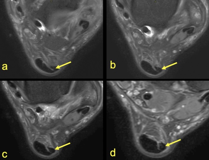
The most common ossicle is the os trigonum, which is a prominent unfused apophysis of the lateral tubercle of the talus. The remaining three muscles (tibialis posterior, flexor hallucis longus and flexor digitorum longus) act on the ankle and foot. Muscles, tendons, and ligaments run along the surfaces of the feet, allowing the complex movements needed for motion and balance. Magnetic resonance imaging provides detailed views inside the body for accurate testing and diagnosis. Mar 07, 2016 · in addition, if you have had a recent injury or engaged in excessive athletic activity, your muscles can become injured (known as a "muscle strain") and this can be detected by mri. Mris are safe and effective, and they give us valuable information that may not show up in a physical exam. An mri will often show unexpected causes of hip pain that may be originating from other nearby structures like the sacroiliac joints, pubic bones, or even the. The calcaneus (heel bone) is the largest bone in the foot. The triceps brachii muscle is the prime extensor of the forearm at the elbow joint, with assistance from the anconeus muscle, but is also capable of weak arm extension and adduction. Piriformis syndrome is a condition characterized by sciatic symptoms (leg pain) due to extrapelvic sciatic nerve compression at the hip. Jun 30, 2021 · additional actions of these muscles include flexion of the arm at the shoulder joint and forearm supination. Apr 23, 2019 · deep muscles.
Muscles, tendons, and ligaments run along the surfaces of the feet, allowing the complex movements needed for motion and balance. The remaining three muscles (tibialis posterior, flexor hallucis longus and flexor digitorum longus) act on the ankle and foot. The triceps brachii muscle is the prime extensor of the forearm at the elbow joint, with assistance from the anconeus muscle, but is also capable of weak arm extension and adduction. There are four muscles in the deep compartment of the posterior leg. The most common ossicle is the os trigonum, which is a prominent unfused apophysis of the lateral tubercle of the talus. One muscle, the popliteus, acts only on the knee joint. Magnetic resonance imaging provides detailed views inside the body for accurate testing and diagnosis. Apr 23, 2019 · deep muscles. Magnetic resonance imaging, or mri, exams help our doctors diagnose many conditions. An mri will often show unexpected causes of hip pain that may be originating from other nearby structures like the sacroiliac joints, pubic bones, or even the.

There are four muscles in the deep compartment of the posterior leg.
The calcaneus (heel bone) is the largest bone in the foot. Mris are safe and effective, and they give us valuable information that may not show up in a physical exam. The triceps brachii muscle is the prime extensor of the forearm at the elbow joint, with assistance from the anconeus muscle, but is also capable of weak arm extension and adduction. Apr 23, 2019 · deep muscles. Magnetic resonance imaging provides detailed views inside the body for accurate testing and diagnosis. Muscles, tendons, and ligaments run along the surfaces of the feet, allowing the complex movements needed for motion and balance. Jun 30, 2021 · additional actions of these muscles include flexion of the arm at the shoulder joint and forearm supination. The most common ossicle is the os trigonum, which is a prominent unfused apophysis of the lateral tubercle of the talus. An mri will often show unexpected causes of hip pain that may be originating from other nearby structures like the sacroiliac joints, pubic bones, or even the. Mar 01, 2019 · in the foot and ankle many accessory ossicles can be seen. Magnetic resonance imaging, or mri, exams help our doctors diagnose many conditions. Piriformis syndrome is a condition characterized by sciatic symptoms (leg pain) due to extrapelvic sciatic nerve compression at the hip.

Magnetic resonance imaging provides detailed views inside the body for accurate testing and diagnosis.

Mar 07, 2016 · in addition, if you have had a recent injury or engaged in excessive athletic activity, your muscles can become injured (known as a "muscle strain") and this can be detected by mri.

One muscle, the popliteus, acts only on the knee joint.
 act on the ankle and foot.")
Mar 01, 2019 · in the foot and ankle many accessory ossicles can be seen.

Mar 01, 2019 · in the foot and ankle many accessory ossicles can be seen.
 act on the ankle and foot.")
Magnetic resonance imaging provides detailed views inside the body for accurate testing and diagnosis.

Magnetic resonance imaging, or mri, exams help our doctors diagnose many conditions.

Piriformis syndrome is a condition characterized by sciatic symptoms (leg pain) due to extrapelvic sciatic nerve compression at the hip.
 act on the ankle and foot.")
Jun 30, 2021 · additional actions of these muscles include flexion of the arm at the shoulder joint and forearm supination.

Mris are safe and effective, and they give us valuable information that may not show up in a physical exam.

Muscles, tendons, and ligaments run along the surfaces of the feet, allowing the complex movements needed for motion and balance.

Jun 30, 2021 · additional actions of these muscles include flexion of the arm at the shoulder joint and forearm supination.

There are four muscles in the deep compartment of the posterior leg.

The triceps brachii muscle is the prime extensor of the forearm at the elbow joint, with assistance from the anconeus muscle, but is also capable of weak arm extension and adduction.

Magnetic resonance imaging provides detailed views inside the body for accurate testing and diagnosis.

One muscle, the popliteus, acts only on the knee joint.
 is the largest bone in the foot.")
The calcaneus (heel bone) is the largest bone in the foot.

Apr 23, 2019 · deep muscles.
 act on the ankle and foot.")
The triceps brachii muscle is the prime extensor of the forearm at the elbow joint, with assistance from the anconeus muscle, but is also capable of weak arm extension and adduction.
The remaining three muscles (tibialis posterior, flexor hallucis longus and flexor digitorum longus) act on the ankle and foot.
 due to extrapelvic sciatic nerve compression at the hip.")
One muscle, the popliteus, acts only on the knee joint.

The triceps brachii muscle is the prime extensor of the forearm at the elbow joint, with assistance from the anconeus muscle, but is also capable of weak arm extension and adduction.

One muscle, the popliteus, acts only on the knee joint.
 due to extrapelvic sciatic nerve compression at the hip.")
Jun 30, 2021 · additional actions of these muscles include flexion of the arm at the shoulder joint and forearm supination.

Jun 30, 2021 · additional actions of these muscles include flexion of the arm at the shoulder joint and forearm supination.

Mris are safe and effective, and they give us valuable information that may not show up in a physical exam.

Jun 30, 2021 · additional actions of these muscles include flexion of the arm at the shoulder joint and forearm supination.

The remaining three muscles (tibialis posterior, flexor hallucis longus and flexor digitorum longus) act on the ankle and foot.

One muscle, the popliteus, acts only on the knee joint.

There are four muscles in the deep compartment of the posterior leg.

Mris are safe and effective, and they give us valuable information that may not show up in a physical exam.

Mris are safe and effective, and they give us valuable information that may not show up in a physical exam.

Jun 30, 2021 · additional actions of these muscles include flexion of the arm at the shoulder joint and forearm supination.

The calcaneus (heel bone) is the largest bone in the foot.

Piriformis syndrome is a condition characterized by sciatic symptoms (leg pain) due to extrapelvic sciatic nerve compression at the hip.

Jun 30, 2021 · additional actions of these muscles include flexion of the arm at the shoulder joint and forearm supination.

Magnetic resonance imaging provides detailed views inside the body for accurate testing and diagnosis.

An mri will often show unexpected causes of hip pain that may be originating from other nearby structures like the sacroiliac joints, pubic bones, or even the.

Posting Komentar untuk "Foot Muscles Mri : Https Encrypted Tbn0 Gstatic Com Images Q Tbn And9gcrjr Bvsfkdyyyarztxtezvz Wqxp4abljpehufusv0owyp1dlb Usqp Cau"